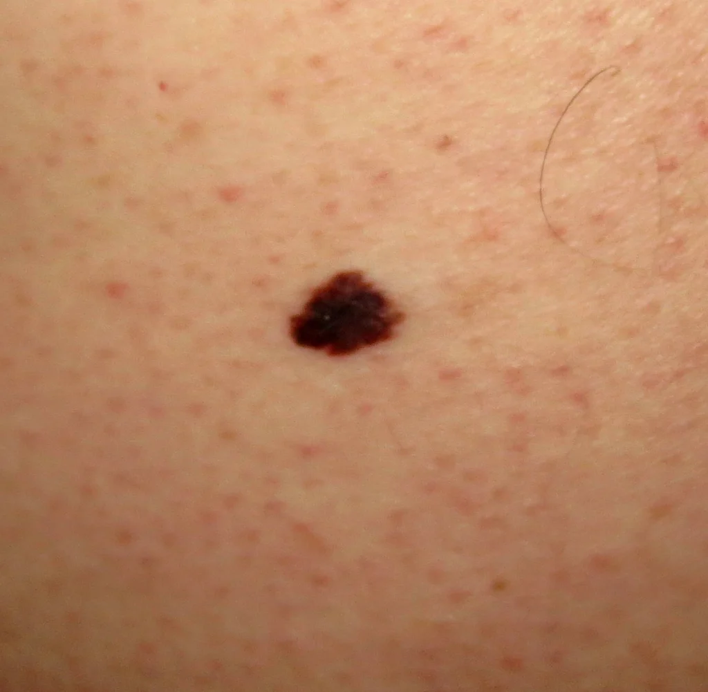
A 45-year-old man comes to the office for a routine preventive examination. He feels well but states that he has always had some dark spots on his back. Physical examination shows many pigmented lesions on the back, including a single, flat, dark, 9-mm lesion with irregular borders. Which of the following is the best next step in the management of this patient's skin lesion?
Below is the code for an example image modal link
Flashcards
/* -- Un-comment the code below to show all parts of question -- */
| A. Excisional biopsy | ||
| B. Incisional biopsy | ||
| C. Photographic documentation and comparison in 6-12 months | ||
| D. Reassurance and routine follow up only |
| Visual assessment of melanoma | |
|---|---|
| ABCDE criteria (≥1 is suspicious) |
|
| 7-point checklist (≥1 major or ≥3 minor is suspicious) |
|
| Ugly duckling sign |
|
This patient’s suspicious pigmented lesion has an increased risk of malignant melanoma based on size (≥6 mm in diameter) and mild border irregularity. Other features that suggest melanoma include asymmetry, color variation, and change over time; some lesions may also have inflammatory changes, crusting/bleeding, or sensory abnormalities.
Due to the aggressive nature and high metastatic potential of melanoma, early referral to a specialist (eg, dermatologist) and definitive diagnosis with biopsy should be considered for any suspicious pigmented skin lesion. Lesion thickness is the most important prognostic determinant in melanoma (ie, increased tumor thickness decreases the survival rate). Therefore, an excisional biopsy with an elliptical technique is the preferred method and should include the entire lesion with a 1- to 3-mm margin. Patients with confirmed melanoma require additional resection and staging procedures, which should be performed within 4-6 weeks of diagnosis.
(Choice B) Incisional biopsy (in which only a portion of the lesion is removed) is not generally recommended for melanoma because it does not allow determination of lesion thickness. However, it may be considered for unusually large tumors or for locations where cosmetic results are important (eg, the face).
(Choices C and D) Periodic surveillance (with or without photographic documentation) is appropriate for patients with lesions that do not require biopsy (eg, symmetric, small) but are at risk for melanoma. This patient’s lesion requires immediate biopsy because of its diameter, evolution over time, and border irregularity.
Educational objective:
Patients with skin lesions suspicious for melanoma should have an excisional biopsy that includes the entire lesion with a 1- to 3-mm margin of surrounding skin and subcutaneous fat.

